news
Celebrating Science Through Imagery
Primary tabs
The Institute for Matter and Systems (IMS) announced the winners of its first-ever IMS Users Image Contest. Selected through public voting, the winning images highlight the remarkable science, innovation, and artistry emerging from research conducted in IMS core facilities.
The contest invited facility users to submit visually compelling images generated through their research—ranging from advanced microscopy and materials characterization to device fabrication and nanoscale analysis. Submissions were showcased in an online gallery, where members of the campus community and broader public were invited to vote for their favorites.
The image contest was created to celebrate the intersection of science and art. Research images not only serve as essential analytical tools, but also reveal patterns, structures, and phenomena that inspire curiosity and creativity.
“We were excited by both the quality of submissions and the level of engagement from the community,” said Eric Vogel, IMS executive director. “These images reflect the extraordinary work happening every day in our facilities and the talent of the researchers who use them.”
The contest also underscores IMS’s commitment to supporting cutting-edge research by providing access to advanced instrumentation, expert staff, and collaborative spaces that enable discovery.
After a community vote, the following winners were identified:
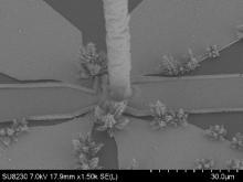
Most Creative | "Carbon Nanotube Candy Cane” By Gabriel Feng

This image was captured on the Hitachi SU8230 SEM in the Materials Characterization Facility. This was a failed overgrown sample from Carbon Nanotube Field Emissions Array.
Most Beautiful | “Snowy Christmas in Nano-ville” By Isha Lodhi
Created using the Hitachi SU8230 in the Materials Characterization Facility.
Most Technically Impressive | “The Lake Monster” By Genaro Soto Valle Angulo
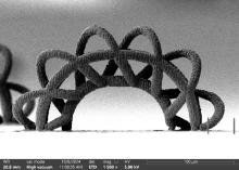
The image was taken on the Thermo Axia SEM. The structures on the image were printed with the Exaddon Metal 3D Printer, out of pure copper with the substrate being a Si wafer coated with a seed layer of copper.
Best Use of Cleanroom Tools| “µCodes: A Universal Grid Platform for Microscale Mapping, Microscopy Navigation, and Multimodal Imaging” By Aref Valipour, part of the Cancer Neurobiology & Nanotechnology Group
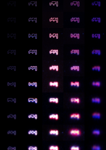
This image was captured on the Olympus MX61 Microscope in the IMS Cleanroom.
Media


Summary
Through its first Users Image Contest, the Institute for Matter and Systems celebrated the creativity and research captured in images produced across its core facilities.
Groups
Status
- Workflow status: Published
- Created by: aneumeister3
- Created: 02/26/2026
- Modified By: aneumeister3
- Modified: 03/04/2026
Categories
Keywords
User Data