news
Digital Dissection: Anatomage Table Brings Anatomy to Life
Primary tabs
Centuries ago, some aspiring doctors resorted to grave robbing to study human anatomy. Today, using the recently purchased Anatomage Table, Georgia Tech students can virtually dissect the human body with a swipe of a touchscreen — no scalpels, no skeletons, and no midnight raids required.
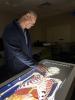
A state-of-the-art anatomy and medical education system, the seven-foot-long Anatomage Table features life-size human — as well as several animal — bodies in digital formats, providing accurate representations of three-dimensional anatomy, physiology, and digital pathology.
“Cadaver dissection is still the gold standard,” explains Senior Academic Professional and Director of Anatomical Sciences Adam Decker, who has taught anatomy and other courses at Georgia Tech since 2010. “But the Anatomage Table lets students interact with living systems digitally — and that’s something we couldn’t offer before.”
Decker is a passionate advocate for using the best tools available to prepare students for medical careers. After leading efforts to bring prosections (pre-dissected specimens that students learn from) to Georgia Tech in 2021, he set his sights on acquiring the Anatomage Table.
“Providing the table was the logical next step,” says Decker. “It’s a way to bridge the tactile experience with dynamic visualization.”
The Anatomage Table was purchased with College of Sciences Technology Fee funds, designed to enhance students' experiences using modern instruments and techniques.
“It’s a great resource for our students, especially for those who are interested in pursuing any field of medicine,” says David Collard, senior associate dean in the College of Sciences. “It supports active learning that will enhance students' applications to medical programs, and gives them experiences with technologies they will encounter in post-graduate professional training.”
Anatomy in action
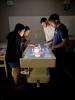
The Series 11 Anatomage Table is housed in the Gilbert Hillhouse Boggs Building and offers a one-to-one display of actual cadavers with five different bodies available for virtual dissection. Students can click on a structure and instantly access detailed information.
“It’s one thing to sit in a classroom and have a professor explain which body parts are which,” says Yusuf Abdalla, a second-year biology student with a pre-med focus. “But being able to independently manipulate the screen to view various parts of the body takes learning to the next level.”
The table offers a cleaner environment with less exposure to odors and chemicals than traditional cadaver dissection.
“Cadavers don’t come with labels. Using the table enables us to see how the body works as a system rather than just viewing individual parts,” adds Rayhan Quraishi, a fourth-year neuroscience major pursuing a career in medicine.
Decker emphasizes that while the Anatomage Table is a game changer, it doesn’t replace prosections. Students will continue to work with real hearts, lungs, and even full spinal cords, thanks to a partnership with Emory University’s Body Donation Program.
Combining cadaver dissection with the table enhances the overall learning experience, explains Decker:
“With prosections, they learn how the veins and arteries feel when you cut into them. With the Anatomage Table, students will see what it looks like when the heart beats or the lungs expand. They can virtually follow a drop of blood through the blood vessel, then use the touch screen to see what that same drop of blood would look like under a microscope. You can’t do that with a cadaver.”
From anatomy to imaging
One of the table’s most powerful features is its integration of diagnostic imaging. Students can compare anatomical structures side-by-side with CT and MRI scans and overlay images as they simulate physiological processes like heartbeats and brain activity.
Decker is currently designing a new course, Anatomy for Diagnostic Imaging, that will use the table to teach students how to interpret MRI, CT, and ultrasound scans. The Anatomage Table contains built-in datasets of MRIs of the spine, heart, and brain, so students can look at the diagnostic image and the actual structure at the same time.
“Some students enter medical school without once taking an anatomy course,” says Decker. “Georgia Tech students, on the other hand, will already have an introduction to imaging and pathology.”
Sameeha Lalani, a third-year biology major who works as an EMT praises the clinical features found in the table. “After one of my EMT shifts, I went back and recreated what happened to my patient using the table. It really made the clinical experience click, so I could better understand what happened.”
Expanding access
The table will soon be in use in BIOS 3754 (Anatomy Lab), which runs five lab sections each fall. Decker is also exploring ways to integrate the table into live lectures, transmitting demonstrations from the table directly into large lecture halls.
Plans are currently underway to use the table in the wellness requirement course, APPH 1040 (Scientific Foundations of Health). Students will be able to visualize cardiovascular anatomy and heart disease by rotating the heart, opening chambers, and simulating conditions, such as a stroke or heart attack.
Decker is eager to collaborate with other departments and make the table a campuswide resource. He sees opportunities in health-related subjects across campus, including biomedical and mechanical engineering, neuroscience, and physiology. Student clubs like the Student Neuroscience Association, Physician Assistant Club, and Pre-Dental Society are also expected to rotate through the lab.
“Anatomy is an ancient science, but it’s the foundation of all healthcare. There are going to be many students who benefit from this — all across campus,” Decker says. “We’ve barely scratched the surface of what it can do.”
What Can Students Do With the Anatomage Table?
- Perform virtual dissections of life-size, digitized human cadavers with touch-responsive controls.
- Rotate, label, and isolate anatomical structures to study systems in detail.
- Compare anatomy with diagnostic imaging, including CT MRI, and ultrasound scans.
- Simulate physiological processes, such as heartbeats, blood flow, and brain activity.
- Explore built-in pathologies, including stroke, tumors, and liver disease.
- Access thousands of annotated structures from male, female, geriatric, pregnant, and animal cadavers.
- Overlay diagnostic images directly onto anatomical models for side-by-side analysis.
- Use real frozen cadaveric slices reconstructed into three-dimensional digital formats.
- Conduct pre- and post-lab activities to reinforce learning before and after cadaver dissection.
- Take anatomy tests, identifying pinned organs and structures.
Media
Summary
Georgia Tech’s new Anatomage Table blends traditional dissection with digital technology — preparing students for the future of medicine.
Status
- Workflow status: Published
- Created by: ls67
- Created: 10/27/2025
- Modified By: ls67
- Modified: 10/30/2025
User Data