event
IEN Imaging Series: Recent Advances and Results in Materials Research and the Life Sciences
Primary tabs
Join us for a technical seminar on recent advances in instrumentation and methods that have opened the door for a variety of new opportunities in 3D characterization and visualization. Specifically, ZEISS X-Ray microscopes (XRM) have uniquely incorporated synchrotron-influenced optical and detection systems to push the boundaries of laboratory XRM, enabling flexible three dimensional imaging capabilities for a wide variety of applications. Similarly, improvements in FIB-SEM instrumentation have enabled new, high resolution applications spanning from nano-patterning and fabrication to 3D imaging and chemical analysis. This talk will cover an overview of XRM and FIB-SEM technology as well as prominent examples and applications, including correlative workflows.
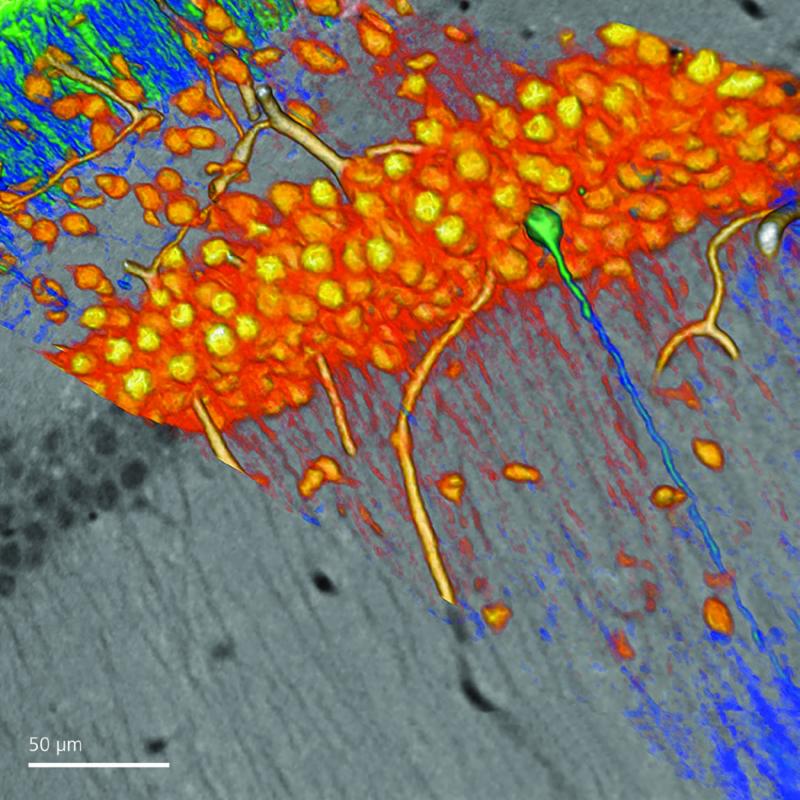
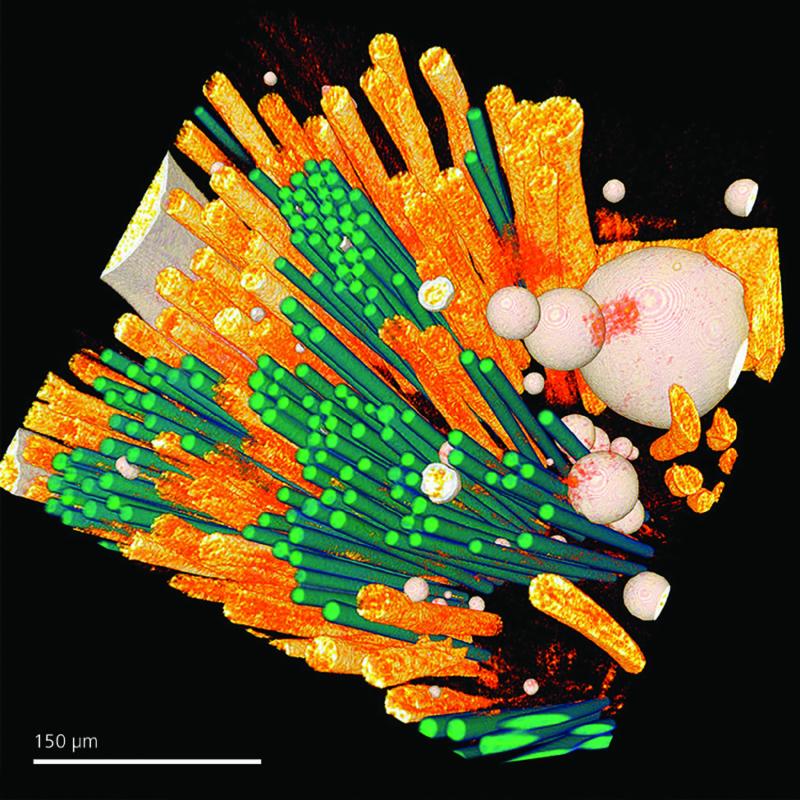 left image: Mammalian brain tissue. Courtesy of the National Center for Microscopy and Imaging Research (NCMIR), University of California, San Diego. Right Image: Non-destructive 3D quantification of fiber reinforced polymer composite materials. Courtesy of Professor Abbas Milani, School of Engineering, University of British Columbia.
For more information about this seminar, or ZEISS imaging, contact Johnafel Crowe
+1 404 536-8477
johnafel.crowe@zeiss.com
Groups
Status
- Workflow status: Published
- Created by: Christa Ernst
- Created: 10/13/2016
- Modified By: Fletcher Moore
- Modified: 04/13/2017
Categories
Keywords
User Data
Target Audience