news
3D Imaging of Transition Metals in the Zebrafish Embryo
Primary tabs
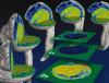
A group of researchers including Chong Shin, an assistant professor in the School of Biology and Christoph Fahrni, a professor in the School of Chemistry and Biochemistry at Georgia Tech conducted a study to visualize the distribution of transition metals in the zebrafish embryo by employing synchrotron X-ray fluorescence (SXRF) microtomography.
By combining the progressive lowering of temperature method (PLT) with femtosecond laser sectioning, a zebrafish embryo at 24 hours post fertilization was embedded, excised, and preserved in an X-ray compatible, transparent resin for tomographic elemental imaging. 3D distribution of zinc, iron, and copper was then reconstructed using the iterative maximum likelihood expectation maximization (MLEM) reconstruction algorithm based on a data set comprised of 60 projections, acquired with a step size of 2 μm during 100 hours of beam time. The volumetric elemental maps, which entail over 124 million individual voxels for each transition metal, demonstrated distinct elemental distributions correlated with characteristic anatomical features at this stage of embryonic development.
The study was published as an ‘inside front cover’ in the journal Metallomics (Bourassa et al., 2014 Sep;6(9):1648-55. doi: 10.1039/c4mt00121d). It has also been selected as one of the outstanding results by the management of the Advanced Photon Source (APS) sector where the work was done.
This project was supported by the National Science Foundation (CHE-1306943), the National Institutes of Health (K01DK081351), and the Vasser Woolley Foundation.
Summary
A group of researchers including Chong Shin, an assistant professor in the School of Biology and Christoph Fahrni, a professor in the School of Chemistry and Biochemistry at Georgia Tech conducted a study to visualize the distribution of transition metals in the zebrafish embryo by employing synchrotron X-ray fluorescence (SXRF) microtomography.
Status
- Workflow status: Published
- Created by: Troy Hilley
- Created: 10/08/2014
- Modified By: Fletcher Moore
- Modified: 10/07/2016
Categories
Keywords
User Data