news
Best Seminar Award Winners
Primary tabs
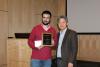
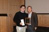
On February 2, 2018, the Best Seminar Award winners in the Wallace H. Coulter Department of Biomedical Engineering at Emory were announced. Participants in the seminar presentation series were evaluated during Fall semester and scored in several areas. Hanjoong Jo, associate chair for Emory and John and Jan Portman Professor in Biomedical Engineering, presented each winner a plaque commemorating their win and a check for $250.
Joan Fernandez Esmerats won in the Ph.D. candidate category. His talk covered, “miR-483 silences UBE2C and prevents aortic valve calcification.”
Seung Yup Lee won in the postdoctoral category. His talk covered, “Non-invasive quantification of brain oxygenation and blood volume in mice and rats using near-infrared spectroscopy.”
Short description of Esmerats talk:
Calcific aortic valve disease (CAVD) is a significant cause of morbidity among the aging population and is a strong risk factor for additional cardiovascular events. Currently, there are no therapeutic options for CAVD other than valve replacement or repair due in part to the incomplete understanding of the underlying mechanisms. Our work has focused in studying a flow-sensitive miRNA, miR-483, that we previously discovered as a potential therapeutic target of CAVD. By conducting an in-silico analysis to discover potential flow-sensitive targets of miR-483 we discovered Ube2c, a key player in the ubiquitin pathway that has never been studied in cardiovascular disease. Our data has showed that miR-483 controls inflammation and calcification via silencing of Ube2c, by treating porcine aortic valves with miR-483 mimic we can significantly decrease aortic valve calcification.
Short description of Lee’s talk:
In brain research, small animal preclinical models provide a valuable platform to study disease mechanisms and to investigate novel therapeutic strategies. Measurements of cerebral oxygen saturation (SO2) and blood volume (CBV) in these animals would aid in the characterization of cerebrovascular-related brain disease progression and therapeutic effects. However, current imaging techniques assessing SO2 and CBV in mice and rats are expensive and typically require a transcranial window, thus limiting their suitability for longitudinal (weeks – months) monitoring. To overcome this limitation, we have developed a novel, non-invasive technique to quantify SO2 and CBV using small-separation frequency-domain near-infrared spectroscopy. We validate this new technique in vitro and in vivo using both tissue-simulating phantoms and neonatal rats. Unlike other techniques, our non-invasive approach provides quantitative measures of resting state oxygen saturation and blood volume without resection of scalp or thinning of the skull of, comparison between animals, longitudinal assessment within a given animal. We plan to employ this method to study preclinical models of sickle cell disease, traumatic brain injury and hypoxic-ischemic encephalopathy.
Status
- Workflow status: Published
- Created by: Walter Rich
- Created: 02/05/2018
- Modified By: Walter Rich
- Modified: 02/07/2018
Keywords
User Data