event
Bioengineering Seminar Series
Primary tabs
“Frontiers in Ultra High Field Magnetic Resonance Imaging: From Brain Function and Connectivity to Angiography in the Human Torso”
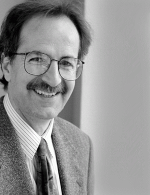
Kamil Ugurbil, PhD
Professor, Director of Center for Magnetic Resonance Research
University of Minnesota School of Medicine
In the last two decades, magnetic resonance imaging (MRI) has evolved as a powerful tool capable of imaging processes such as organ function, intracellular chemistry, tissue perfusion, oxygen utilization, and enzyme activity in intact animals and humans. Most notable among these capabilities is functional magnetic resonance imaging (fMRI), which, in 2012, celebrates its twentieth year since its discovery. During these two decades, we have systematically pursued ever-increasing magnetic fields to improve the information content available from MRI studies, going first to 4 Tesla, and subsequently to 7 and 9.4 Tesla for human imaging. A plethora of early experiments, particularly at 7 Tesla, demonstrated superior sensitivity and accuracy of fMRI signals, and improvements in several contrast mechanisms for anatomical and physiological imaging. In fMRI, these gains have ultimately resulted in unique applications such as functional mapping of elementary computational units in the human brain (orientation domains in V1 and direction selective organizations in human MT), and visualization of functional brain networks with unprecedented spatial and temporal detail. These applications involved numerous engineering solutions and methodological developments to challenges posed by high magnetic field MRI, including the complexities arising from damped traveling wave behavior of 300 MHz RF, the 7T proton frequency, in the human body. With these engineering and methodological solutions, human imaging at 7T and higher fields has been feasible not only in the human head but also in the human torso, where the challenges have been considered insurmountable until our recent work, leading to accomplishments such as coronary and kidney angiography without contrast agents and morphological imaging of the torso in high resolution and contrast.
Status
- Workflow status: Published
- Created by: Colly Mitchell
- Created: 11/16/2011
- Modified By: Fletcher Moore
- Modified: 10/07/2016
Categories
Keywords


