news
Visualizing the Itch-Sensing Skin
Primary tabs
Chronic itch is defined as itch persisting for more than six weeks. Because chronic itch is associated with most skin diseases, it is the most common reason for visiting a dermatologist. In addition to being uncomfortable, repeated scratching may result in infection and scarring, making chronic itch socially and occupationally debilitating.
Until recently researchers have experienced difficulty in visualizing the itch-sensing neurons that innervate the skin and are responsible for sensing itch sensation. However, a team of Georgia Tech researchers from the School of Biological Science has combined different cutting-edge techniques to solve this problem.
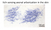
“We created a new transgenic mouse line that allowed us to, for the first time, see individual itch neurons in the skin,” says Yanyan Xing, a postdoctoral fellow in the Han laboratory. “This is very exciting!” she continued, “Because there are so many neurons in the skin, they often overlap on top of one another. This makes it impossible to determine the size, frequency, or distribution of the neurons.”
Such a state makes it impossible for researchers to perform any sort of detailed analysis on the neurons. For instance, the researchers cannot tell the number of axons per neuron, look for patterns in the spatial density of neurons, or see if the neurons are attached to any specific structures. “In contrast,” Xing explained, “our transgenic mouse line allows us to perform ‘sparse-labeling’ so that only a few neurons, less than 1%, are visible. Now, we can visualize individual neurons!”
Xing completed this work with a graduate student, Haley Steele, and four other fellow School of Biological Sciences researchers under the direction of Dr. Liang Han. The team published their results, “Visualizing the Itch-Sensing Skin Arbors,” in The Journal of Investigative Dermatology. Specifically, the team looked at a group of itch sensing neurons that are identified by the presence of a single protein, MrgprC11. They, therefore, call this group of neurons MrgprC11+ itch-sensing neurons.
To visualize these MrgprC11+ neurons, the team used a histological staining technique known as PLAP. This technique turns the individual axons of the neurons a dark blue which is visible to the naked eye, even without the use of a microscope.
By visualizing the individual neurons, the team discovered that itch-sensing neurons have large receptive fields. “Receptive fields are the area on the skin that each neuron is responsible for sensing,” Xing explains. “So, if the receptive field is small, such as for touch, you can sense very precisely that something is touching you at this very particular spot. But for the MrgprC11+ itch neurons, we found that they had large receptive fields, three times bigger than for the other neurons we looked at. So that means that when we sense itch, it isn’t confined to a very particular spot. We feel it much more diffusely over a larger area.”
In addition to allowing for the visualization of the itch neurons in the skin, this team’s novel transgenic mouse line also allowed them to learn more about MrgprC11+ neurons in general. For example, they discovered that MrgprC11+ neurons have multiple itch receptors. This is a critical finding according to Xing because “previously nobody was really looking too closely at the MrgprC11+ neurons. Now, that we know that MrgprC11+ neurons are an important itch sensing neuronal population, future researchers may focus significantly more effort on studying MrgprC11+ neurons.”


Status
- Workflow Status:Published
- Created By:Jasmine Martin
- Created:01/15/2021
- Modified By:Jasmine Martin
- Modified:01/15/2021
Categories
Keywords