news
Petit Institute Adds New Super-Res Microscope to Core Facilities
Primary tabs
Researchers in the Parker H. Petit Institute for Bioengineering and Bioscience are developing a clearer picture of biological inner space thanks to a new super-resolution fluorescence microscope, the newest piece of equipment in the institute’s core facilities universe.
“I call it the million dollar microscope,” says Steve Woodard, core facilities manager at the Petit Institute. “This is not the kind of equipment you’re going to find on just any university campus.”
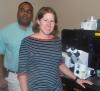
The versatile Zeiss Elyra PS. 1 microscope was installed in the Petit Biotechnology building in late February, and it has allowed student and faculty researchers a better view of unseen elements of life.
“My lab has already used it to resolve how nanoparticles cluster on the cell surface, which is really neat because normally, nanoparticles are too small to resolve,” says Christine Payne, Petit Institute faculty member and associate professor in the School of Chemistry and Biochemistry.
Getting a closer, clearer look at how nanoparticles interact with cells could lead to the design of improved drug delivery systems, says Payne, who serves on the Core Facilities Advisory Committee and suggested to the group that the Petit Institute could really benefit from the state-of-the-art microscope.
They readily agreed, and she took the lead in submitting the grant proposal that secured $469,000 through the National Science Foundation’s Major Research Instrumentation (MRI) Program, designed to increase access to shared scientific and engineering equipment for universities and other non-profit research centers.
“You see, here was a researcher who identified a need for facilities, and had the backing and support of the Petit Institute,” says Woodard with the air of someone who has seen this happen before, because he has.
Woodard has seen core facilities grow all around him in the nearly 20 years he’s been with the Petit Institute, from one confocal microscope in a single room to $15 million of equipment – 50 pieces scattered over 3,000 feet of unconnected space, and this latest piece arrived in similar fashion to all the other stuff that came before it.
“It really took a group effort, in every sense,” Woodard says.
For one thing, according to Payne, about 25 people were involved in the proposal to get the microscope, including the Petit Institute’s grand administrator, Rachel Cochran. But the team approached worked especially well when it came down to counting pennies.
NSF granted almost half a million, and Woodward says, “we were still short, but the College of Engineering and the College of Sciences, as well as the Petit Institute, really stepped up the plate and made it happen.”
So did the School of Chemistry and Biochemistry, the School of Biology, and the School of Physics. That collective largesse brings to the Petit Institute a versatile microscope, designed to take imaging beyond the diffraction limit of standard confocal microscopy, utilizing either structured illumination microscopy (SIM) to increase the resolving capabilities down to about 100 nm (nanometers), or photo activation light microscopy (PALM), to resolve down to 20 nm.
The main advantage of fluorescence microscopy, as opposed to something like electron microscopy, is its compatibility with living cells.
“This microscope uses what I’d call optical tricks and specialized image processing systems to get you down to an amazing level of resolution, beyond the normal resolution of visible light,” Payne says. “One of the advantages of that is, we can use live cells. And that’s a big deal because we like to use live samples.”
Her grad students apparently like the new microscope so much they invited the co-developer of the first super-high-resolution PALM microscope, Eric Betzig, to be their speaker at the Peter B. Sherry Lecture (Thursday and Friday, April 24-25). It’s an annual event hosted by the School of Chemistry and Biochemistry’s Graduate Student Forum.
In Thursday night’s opening lecture, Betzig, whose lab develops optical imaging tools at Janelia Farm (part of the Howard Hughes Medical Institute outside Washington, D.C.), gave a rundown of his history in the field, and where Janelia’s groundbreaking research is headed, to the approximately 100 students and faculty in attendance.
Betzig, one of the innovators in his field, spoke frankly and humorously (and occasionally bluntly) about advancements in microscopy. Bottom line: There’s lots of inner space left to explore.|
“The good news is,” Betzig says, “that the standard tools biologists use to study live cells leave a lot of room for improvement.”
Media

Summary
Microscope will give closer, clearer look at how nano particles interacts with cells
Status
- Workflow status: Published
- Created by: Colly Mitchell
- Created: 04/28/2014
- Modified By: Fletcher Moore
- Modified: 10/07/2016
Keywords
User Data