image
Cell activity
Primary tabs

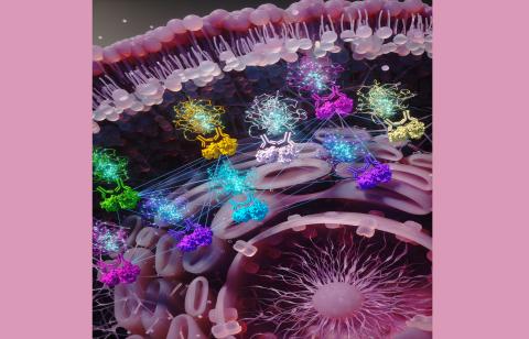
An artistic rendering of sub-cellular activity: The cell membrane is seen at the top, nucleus on the bottom/right. Protein pairs are being targeted by antibodies (sets of two). Then antibodies are linked to DNA pieces that glow when proteins were found to be closely interacting with each other. The glowing fluorescence DNA signal is then imaged by a microscope indicating the spatial locations of protein interactions as dots, which researchers use to generate graph models. The straight lines connecting the antibody and protein pairs indicate their graph wiring that gets altered in drug resistance.
