news
Georgia Tech Researchers Seek a Better Understanding of the Brain
Primary tabs
When you look at a color, hear a sound or smell a favorite aroma, what part of your brain goes into action? When you drive a car or recognize a face, which part of your brain comes alive with the electrical impulses of firing neurons? If your brain is injured, how does it work differently?
Scientists and engineers at the Georgia Institute of Technology are applying their expertise, tools and techniques to address questions like these – and to explore on a fundamental level how the brain works.
Because the human brain is immensely complex, the researchers are pursuing many levels of inquiry – from molecules to cells to circuits to the mystery of the mind itself – and also studying brain disorders and development, along with daily feats of brain activity, such as vision, speech, movement and memory.
Georgia Tech researchers are also developing better interventions for brain injuries and disorders. They are designing tools to help neuroscientists better probe and record the activity of neurons in tissue samples and living animals. And they are using brain imaging techniques, such as magnetic resonance imaging (MRI) and electroencephalography (EEG), to peek inside the skull and examine how the brain reacts differently when cognitive tasks are completed by the young and the old, or the healthy and those with injuries.
This article provides a snapshot of Georgia Tech’s research in the biology of the brain.
Developing Better Interventions for Brain Disorders and Injuries
Reducing Epileptic Seizures -- Researchers at Georgia Tech and Emory University are investigating the use of electrical stimulation to reduce or eliminate seizures associated with epilepsy, a disorder that affects approximately 2 million people in the United States. Seizures are temporary disturbances in brain function in which groups of nerve cells in the brain fire abnormally and excessively.
To perform their studies, the researchers have created an animal model for temporal lobe epilepsy. Using this model, they can examine different approaches for preventing seizures associated with epilepsy. For one approach, they are implanting tiny electrodes in the animal’s brain that can be used to stimulate neurons and record their activity. The team is also trying to utilize the field of optogenetics – a mix of optical and genetic techniques – to stop the seizures by stimulating the brain with light.
“Our goal is to better understand what causes epileptic seizures and try to find a way to respond to those bursts in activity with stimulation and reduce the number of seizures an individual experiences,” said Steve Potter, an associate professor in the Wallace H. Coulter Department of Biomedical Engineering at Georgia Tech and Emory University.
The stimulation techniques could be a possible alternative for individuals who do not respond to drug therapies and may therefore require surgical resection of the portion of the brain causing the seizures.
Potter is collaborating on this project with Robert Gross, an associate professor in the Departments of Neurosurgery and Neurology at Emory University, and a member of the program faculty in the Coulter Department. Their graduate students, Sharanya Desai and Neal Laxpati, are developing and testing these new brain stimulation therapies in the epileptic rat model. This work has been funded in part by the Wallace H. Coulter Foundation, the National Institutes of Health, Citizens United for Research in Epilepsy (CURE) and the American Epilepsy Society.
Improving Recovery from Spinal Cord Injuries -- Following an injury to the brain or spinal cord, a glial scar begins to form. While the scar signifies the beginning of the healing process, neuron extensions – called axons – cannot regenerate through the glial scar, thus preventing repair and recovery.
The inhibitory characteristics of the scar have been attributed to an increase in proteins known as chondroitin sulfate proteoglycans at the injury site. This family of proteins prevents regeneration of damaged nerve endings.
In a recent study, a research team led by Ravi Bellamkonda, a professor in the Wallace H. Coulter Department of Biomedical Engineering at Georgia Tech and Emory University, examined the influence on central nervous system recovery of a chondroitin sulfate proteoglycan called chondroitin sulfate-4,6 (CS-E). The researchers found that expression of CS-E increased following a central nervous system injury. In cell culture experiments, CS-E inhibited the growth of neurons, and when researchers reduced the amount of CS-E, the inhibition of neuron growth was significantly alleviated.
“Our findings showed that CS-E is a big player in inhibiting nerve growth following an injury, and its expression needs to be reduced as much as possible,” said Bellamkonda.
One strategy to overcome the inhibitory effects of proteins like chondroitin sulfate-4,6 is to enzymatically digest them. In 2009, Bellamkonda developed an improved version of an enzyme capable of digesting chondroitin sulfate proteoglycans.
The researchers eliminated the thermal sensitivity of the enzyme – called chrondroitinase ABC (chABC) – and developed a delivery system that allowed the enzyme to be active for weeks without implanted catheters and pumps. In animal studies, when the thermostabilized enzyme was delivered, the scar at the injury site was significantly degraded for at least six weeks, and enhanced axonal sprouting and recovery of nerve function at the injury site were observed.
“These results brought us a step closer to repairing spinal cord injuries, which require multiple steps including minimizing the extent of secondary injury, bridging the lesion, overcoming inhibition due to scar, and stimulating nerve growth,” said Bellamkonda, who is also the Carol Ann and David D. Flanagan Chair in Biomedical Engineering and a Georgia Cancer Coalition Distinguished Cancer Scholar.
Robert McKeon, an associate professor in cell biology at Emory University, Georgia Tech senior research scientist Lohitash Karumbaiah and graduate student Hyun-Jung Lee also contributed to this work, which was supported by the National Institutes of Health and the Wallace H. Coulter Foundation.
Uncovering the Neural Basis of Rapid Brain Adaptation -- Your brain is able to quickly switch from detecting an object flying toward you to determining what the object is through a phenomenon called adaptation.
Garrett Stanley, an associate professor in the Wallace H. Coulter Department of Biomedical Engineering at Georgia Tech and Emory University, published a study in the journal Nature Neuroscience that detailed the biological basis for rapid adaptation: neurons located at the beginning of the brain’s sensory information pathway that change their level of simultaneous firing. This modification in neuron firing alters the nature of the information being relayed, which enhances the brain’s ability to discriminate between different sensations – at the expense of degrading its ability to detect the sensations themselves.
“Previous studies have focused on how brain adaptation influences how much information from the outside world is being transmitted by the thalamus to the cortex, but we showed that it is also important to focus on what information is being transmitted,” said Stanley.
Recording how neurons in different parts of the brain simultaneously communicate with each other in different situations is a big step in the neuroscience field. The researchers plan to use the techniques from this study to probe the effects of brain injury, which can change the degree of synchronization of neurons in the brain, resulting in harmful effects.
In addition to Stanley, Coulter Department research scientist Qi Wang and Harvard University researchers contributed to this work, which is supported by the National Institutes of Health.
Filling the Neuroscience Toolbox
Device for Probing Neurons in Tissue Samples -- Axion BioSystems, a startup company based on intellectual property developed at Georgia Tech, offers neural interfacing technologies for basic science, and for pharmaceutical and clinical research applications. The company has developed microelectrode arrays (MEAs) that allow simultaneous stimulation and recording of neural tissue, and include low-power chips that can service hundreds of channels.
“Our objective has been to develop devices that can precisely manipulate and monitor electrically active cells and tissues of many types – including brain, spinal, muscle and cardiac – and provide real-time access to complex electrophysiological information,” said James Ross, the company’s chief technical officer. “Researchers using Axion’s technology capture biological models of human heartbeats and brain waves in a dish, which opens the door to a wide range of drug development and safety tests.”
In addition to Ross and company CEO Tom O’Brien, Axion BioSystems was founded by School of Electrical and Computer Engineering professor Mark Allen, Department of Biomedical Engineering professor Stephen DeWeerth, research engineer Edgar Brown and Swami Rajaraman, a recent Ph.D. graduate.
Axion has raised more than $9 million from private investors, grants from the National Institutes of Health’s Small Business Innovation Research (SBIR) program and early-stage funding from the Georgia Research Alliance (GRA). The company resides in laboratory and office space at the Advanced Technology Development Center (ATDC) biosciences incubator on Georgia Tech’s campus.
The company is currently working to increase the sales and adoption of its products by pharmaceutical companies, contract research organizations and academic institutions. Since it was founded in 2007, the company has grown from two to 20 employees and launched two commercial products – the Muse and the Maestro.
“The technology we licensed from the Georgia Tech Research Corporation allows us to provide two MEA systems that reduce the cost and complexity of conducting neuroscience research,” explained Ross. “Both systems consist of low-cost, disposable multielectrode arrays, and integrated circuits that eliminate stimulation artifacts and enable simultaneous stimulation and recording.”
The Muse is a bench-top system containing 64 channels for stimulating and recording electroactive tissue. The high-throughput Maestro contains 768 stimulating and recording channels, accommodates multiwell plates of up to 96 wells and is suited for large-scale cellular analysis in commercial drug screening applications.
While the company’s current efforts are focused on pharmaceutical drug screening, ongoing development is expected to result in products in the medical diagnostic and medical device arenas, Ross said.
Devices for Probing Neurons in Living Animals -- When high-fidelity recording of individual neurons in live animals is required, whole-cell patch clamp electrophysiology of neurons in vivo is the gold-standard, but it requires great skill to perform. The technique utilizes a glass micropipette to establish electrical and molecular connections to the insides of neurons embedded in intact tissue to record synaptic and ion-channel-mediated events.
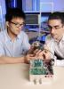
Researchers at Georgia Tech and the Massachusetts Institute of Technology (MIT) have developed a simple robot that automatically performs whole-cell patch clamping in vivo. Using the robot, the researchers have demonstrated high throughput and recording quality in the cortex and hippocampus of small animals.
“With the robot, neuroscientists can achieve high-quality recordings with yields that exceed those of skilled humans at speeds sufficient to enable an unskilled human operator to clamp dozens of cells or more per day and collect data about each one’s gene expression, shape and electrical behavior,” said Craig Forest, an assistant professor in the George W. Woodruff School of Mechanical Engineering.
Applications for the autopatching robot include studying the effects of drugs on neuron electrophysiology; examining neuron behavior in disease states, such as epilepsy and narcolepsy; and classifying neuron cell types on a high-throughput scale.
The robot was designed by Forest; Georgia Tech graduate student Suhasa Kodandaramaiah; Edward Boyden, an associate professor of biological engineering and brain and cognitive sciences at the MIT Media Lab and MIT McGovern Institute; MIT graduate student Giovanni Franzesi; and MIT postdoctoral researcher Brian Chow.
The researchers recently created a startup company, Neuromatic Devices, to commercialize the device. Development of the new technology was funded primarily by the National Institutes of Health, the National Science Foundation and the MIT Media Lab.
Maysam Ghovanloo, an associate professor in Georgia Tech’s School of Electrical and Computer Engineering, has developed a wireless system that collects neural signals from awake, freely moving animals during behavioral neuroscience research experiments. The Wireless Implantable Neural Recording (WINeR) system can simultaneously record from 32 channels for an unlimited period of time using a wireless inductive power transmission system.
“The WINeR system removes the need to tether a small animal via cable to a neural recording device during behavioral neuroscience research experiments and relieves the animal from carrying bulky batteries, thus eliminating two major sources of motion artifacts and bias,” said Ghovanloo.
WINeR is powered by the EnerCage system, which consists of an array of overlapping spiral planar coils that cover the bottom of the experimental area and enable inductive power transmission. A mobile unit attaches to the animal to regulate and deliver a constant amount of inductive power to the WINeR device and any other electrophysiology sensors used to collect data during an experiment, despite animal movements. The mobile unit also contains a small magnet that allows the animal’s location to be tracked in real time.
The researchers plan to add the functionality of wirelessly stimulating neurons to the WINeR device and increase the number of channels it provides.
Ghovanloo is collaborating with Joseph Manns, an assistant professor in the Emory University Department of Psychology, and Karim Oweiss, an associate professor in the Michigan State University Department of Electrical and Computer Engineering and the Neuroscience Program, to test the WINeR and EnerCage systems. This work is supported by the National Science Foundation and the National Institutes of Health.
To alleviate the need for electrodes implanted in the brain, researchers in the Georgia Tech Research Institute (GTRI) are collaborating with Neural Signals Inc. to explore the potential use of near-infrared fluorescent probes to wirelessly transmit neural signals from inside the brain to an external recording device.
A team led by GTRI principal research scientist Brent Wagner is investigating the possibility of connecting neurons to a wireless neural interface system that could respond to low-voltage, low-frequency electrical signals in the brain. The system would consist of a grid of gold nanoparticles, each linked via flexible strand of DNA to a semiconductor quantum dot.
With this system, when a neural cell is at rest, the quantum dot and gold nanoparticle are in close proximity, so no light is emitted from the quantum dot. When a neural cell fires, the voltage change on the neuron’s surface pushes the quantum dot away from the gold nanoparticle, allowing the quantum dot to emit light. The precise location of the quantum dot’s near-infrared luminescence can be detected using an infrared camera.
“The sensing mechanism for the system is based on energy transfer between the quantum dot and the gold nanoparticle,” said Wagner. “We think one of the major advantages of this type of system is its potential to transmit a high throughput of neural signals from multiple recording sites at the same time without the use of bulky cables or implanted electrodes.”
This project is supported by the GTRI Independent Research and Development (IRAD) program.
Researchers in the Georgia Tech School of Chemical and Biomolecular Engineering are building devices to help neuroscientists better understand how neurons in the brain contribute to an organism’s behavior.
Using inexpensive components from ordinary LCD projectors, associate professor Hang Lu can control the brain and muscles of freely moving tiny organisms, including the Caenorhabditis elegans worm that is commonly used for biological studies. Red, green and blue lights from the projector activate light-sensitive microbial proteins that are genetically engineered into the worms, allowing the researchers to switch neurons on and off like light bulbs and turn muscles on and off like engines.
The inexpensive illumination technology allows researchers to stimulate and silence specific neurons and muscles of the worms, while precisely controlling the location, duration, frequency and intensity of the light.
Use of the LCD technology to control small animals advances the field of optogenetics – a mix of optical and genetic techniques that has given researchers unparalleled control over brain circuits in laboratory animals. Until now, the technique could be used only with larger animals by placement of an optical fiber into an animal’s brain, or by illumination of an animal’s entire body.
For another project, Lu developed a microfluidic device that enables genetic studies on small organisms to be performed more quickly. An addition to the system since its original development is a laser beam that can destroy individual neurons. By monitoring the animal’s behavior after the laser ablation, the researchers can infer the function of each neuron. The process takes only 20 to 30 seconds, much less than the half hour it can take to ablate neurons using other techniques.
Lu and collaborators at the Queensland Brain Institute and the University of Queensland in Brisbane, Australia, have also adapted the original design of the microfluidic device to a curved geometry that enables positioning C. elegans bodies into lateral orientations. This alignment makes it easier to analyze neuronal developmental and disease processes that travel from the worm’s head to end or laterally across the worm’s body. Results of this research were published in April 2012 in the journal PLoS ONE.
“These systems have many applications in developmental and behavioral neuroscience of model organisms,” said Lu. “Our challenge is to make them as easy to use as possible so that the technology can make an impact in biological and medical research.”
Lu’s research is supported by the National Science Foundation, the National Institutes of Health and the Alfred P. Sloan Foundation.
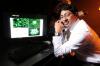
Models of How the Brain Processes Information -- Christopher Rozell, an assistant professor in the Georgia Tech School of Electrical and Computer Engineering, uses mathematical models and signal processing technologies to understand how the brain organizes and processes images and sounds.
“Machine systems and the human brain perform similar tasks, such as speech recognition and computer vision, but the machines still fall far short of the human brain in these tasks, especially in the areas of power consumption and efficiency,” said Rozell.
In the brain, information about a stimulus in the outside world is communicated to higher centers in the brain by a collection of electrochemical signals present in groups of neurons. Recent evidence indicates that these groups of neurons may represent information by activating only a few of these units – known as a sparse code – and never centralizing the information in a single decision-making unit.
While sparse coding in neural systems is not well understood, Rozell and School of Electrical and Computer Engineering professor Jennifer Hasler and associate professor Justin Romberg are developing neurally plausible analog circuits to quickly find sparse codes. This approach could potentially solve problems relevant for many engineering applications much faster, while using less power than a traditional digital system.
“We don’t have the time or capability to record the characteristics and properties of each of the billions of neurons in the brain to validate our models, but we know our models of neural coding for sensory information are biophysically realistic because we verify them against published results of electrophysiology experiments,” said Rozell.
Researchers in Rozell’s laboratory are also examining what advantages a sparse code might have for the brain, which is making perceptual judgments based on visual data. By investigating how the brain transforms the outside world into meaningful representations it can work with, Rozell hopes better brain-machine interfaces can be designed, more efficient signal processing systems can be developed, and vision and hearing deficits can be corrected. This research is supported by the National Science Foundation and the National Institutes of Health.
Monitoring Activity in the Brain During Cognitive Tasks
Picking Out the Right Tool -- Choosing how to use tools to accomplish a task is a natural and seemingly trivial aspect of our lives, yet it can be very difficult for persons with certain brain injuries.
“In my laboratory, I study cognitive motor control,” said Georgia Tech School of Applied Physiology assistant professor Lewis Wheaton. “I want to understand the neural system that allows us to select the best tool to accomplish a task, pick that tool up and use it correctly to complete the task without overloading our brains with information.”
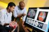
In a recent study, Wheaton identified neural activation patterns in the brain associated with watching tools used in correct and incorrect contexts. He used the functional MRI (fMRI) scanner at the Georgia State/Georgia Tech Center for Advanced Brain Imaging, along with electroencephalography (EEG), to record neural activations in the brain as healthy individuals identified whether tools shown in photographs were being used in correct or incorrect contexts. For example, a participant might be shown a hammer and nail, which is a correct tool use, or a hammer and coffee mug – an incorrect tool use.
The fMRI results revealed that when participants identified correct tool use, different parts of the brain became active compared to when they identified incorrect tool use. The EEG recordings provided additional information about the evolution of these activations over time. Activation occurred between 300 and 400 milliseconds after a correct tool use image was shown, but more quickly following onset of an incorrect tool use image. These findings were published in the journals Brain Research and Frontiers in Human Neuroscience.
Wheaton is now using the information he learned about the neural mechanisms of tool use in healthy brains to better understand tool learning and why some individuals experience impaired tool-related behavior following a stroke – a deficit called apraxia.
“In conceptual apraxia, we think the network that codes for incorrect tool use may be selectively damaged and incorrect contextual information is being passed to the areas of the brain activated by correct tool use. Because no error signal arises, contextually inappropriate use becomes possible,” said Wheaton.
Predicting an Individual’s Attentiveness -- Shella Keilholz’s long-term research goal is to build a model of spontaneous activity in the brain. As an engineer, she views the brain as a collection of hierarchical networks, with local networks of cells that work together and larger networks where information is transferred between different areas in the brain.
Keilholz is part of a team that is using the fMRI scanner at the Georgia State/Georgia Tech Center for Advanced Brain Imaging to probe the functional connectivity of the brain while an individual is performing a cognitive task requiring vigilance. The researchers are investigating whether the complex neural interactions between spatially distinct brain regions can be used to predict how well an individual will perform on cognitive tasks.
Funding for this work is provided in part by the U.S. Air Force through the Bio-nano-enabled Inorganic/Organic Nanostructures and Improved Cognition (BIONIC) Air Force Center of Excellence at Georgia Tech.
The team’s goal is to find a stable marker in the fMRI signal that is associated with cognitive processing and alertness. Initial results of their experiments show that the level of brain activity preceding the presentation of a visual stimulus can predict how fast an individual will respond to the stimulus during a vigilance task.
“U.S. Air Force analysts must remain attentive to computers and controls for hours at a time, so we are trying to develop a noninvasive way to measure the current state of an individual’s brain and determine if that person is getting off task,” said Keilholz, an assistant professor in the Wallace H. Coulter Department of Biomedical Engineering at Georgia Tech and Emory University. “With that information, one might be able to develop a way to refocus that person and get him or her back on task, which would optimize work effectiveness and possibly save lives.”
Also contributing to this project are School of Psychology associate professor Eric Schumacher, and Air Force Research Laboratory biomedical engineer Andrew McKinley and integration manager Lloyd Tripp.
Recalling Memories -- Audrey Duarte, an assistant professor in Georgia Tech’s School of Psychology, is a cognitive neuroscientist – someone who looks at the neuroscience that supports human behavior. Duarte’s research is focused on episodic memory, which is the memory of specific events, situations and experiences. Your first day of school, attending a friend’s birthday party and what you ate for dinner last night are examples of episodic memories.
Episodic memory can be affected by a number of disorders – including stroke, dementia and Alzheimer’s disease – and even healthy aging. Through her research, Duarte is trying to understand what happens as the brain ages to cause decline in memory abilities over time.
“We want to determine if there are specific areas in the brain or specific brain networks that are disproportionately affected in a negative way by aging, causing lapses in episodic memory,” she said.
Using the fMRI scanner at the Georgia State/Georgia Tech Center for Advanced Brain Imaging, Duarte measures activity from thousands of neurons in the brain at the same time and assesses the patterns of activity while young and older adults examine and subsequently retrieve pictures of common objects from memory. Using this data, Duarte is developing strategies to help older adults better encode and retrieve episodic memories.
“By finding out where an individual’s attention is drawn when looking at a picture, we can better understand the relationship between attention and memory and look for ways to remediate impairments in episodic memory,” said Duarte.
This research is supported by the National Science Foundation, the National Institutes of Health and the American Federation for Aging Research.
Accomplishing Fine Motor Tasks -- In another project, Georgia Tech researchers are studying the effects of aging on the neural connectivity between the motor cortex and muscles during tasks that require fine motor skills.
“We know that aging and dual-task paradigms often degrade fine motor performance, so we wanted to compare the performance of young and older adults during the execution of a fine motor task alone and concurrent tasks that required substantial divided attention,” said Minoru Shinohara, an associate professor in the Georgia Tech School of Applied Physiology.
For the study, two groups of healthy adults, one group between the ages of 18 and 38 and the other between 61 and 75, performed tasks involving one-finger motor, two-finger motor, cognitive and concurrent motor-cognitive skills.
As the participants completed the tasks, Shinohara and School of Electrical and Computer Engineering graduate student Ashley Johnson examined the synchrony between two signals – an electroencephalogram (EEG) acquired from the primary motor cortex in the brain and an electromyogram (EMG) acquired from a muscle in the hands. The synchronous measurement is called corticomuscular coherence.
In the study, the older adults demonstrated higher corticomuscular coherence than the young adults during performance of both unilateral and dual tasks. Corticomuscular coherence was highest in the older adults, especially during the dual motor-cognitive task and increased with an additional task for both groups of subjects. But during the motor-cognitive task, corticomuscular coherence was negatively correlated with motor output error across young, but not older, adults. The results of the study were published online in January 2012 in the Journal of Applied Physiology.
“The findings demonstrate that older and younger adults don’t need to use the same neural strategy to accomplish the same motor performance,” said Shinohara. “We are seeing changes in neural strategies for accomplishing fine motor skills with aging.”
In addition to aging, these types of changes in neural strategies could be valuable for rehabilitation applications. Individuals with neurological deficits might benefit from using a different strategy to perform motor tasks, rather than using the same strategy they used before the deficit occurred.
Georgia Tech’s extensive involvement in neuroscience research – from basic to clinical science – reflects the interests of researchers from multiple academic departments and the Georgia Tech Research Institute (GTRI). The researchers are working to better understand how the brain works and apply this knowledge to improving brain function, which has applications for those who have sustained losses due to injuries or disease.
Research reported in this publication was supported by the National Institute of Neurological Disorders and Stroke (NS054809, NS079268, NS043486, NS48285, NS062031 and NS058465), the National Institute of Biomedical Imaging and Bioengineering (EB009437 and EB012803), the National Institute on Aging (AG035317 and AG016201), the National Institute of General Medical Sciences (GM088333), the National Eye Institute (EY019965), the National Science Foundation (ECCS-0824199, CBET-0954578, DBI-0649833, CCF-0905346 and BCS-1125683), and the U.S. Air Force (FA9550-09-1-0162). The content is solely the responsibility of the principal investigators and does not necessarily represent the official views of the NIH, NSF or U.S. Air Force.
Research Horizons Magazine
Georgia Institute of Technology
177 North Avenue
Atlanta, Georgia 30332-0181 USA
Media Relations Contact: John Toon (404-894-6986)(jtoon@gatech.edu)
Writer: Abby Robinson
Media


Summary
Scientists and engineers at Georgia Tech are applying their expertise, tools and techniques to explore on a fundamental level how the brain works. Because the human brain is immensely complex, the researchers are pursuing many levels of inquiry
Groups
Status
- Workflow Status:Published
- Created By:John Toon
- Created:06/24/2013
- Modified By:Fletcher Moore
- Modified:10/07/2016
Categories
Keywords